Scientists Reconstruct the Face of the Oldest Known Homo Sapiens
A digital facial approximation built from 315,000-year-old Moroccan fossils reveals what the earliest known Homo sapiens may have looked like.
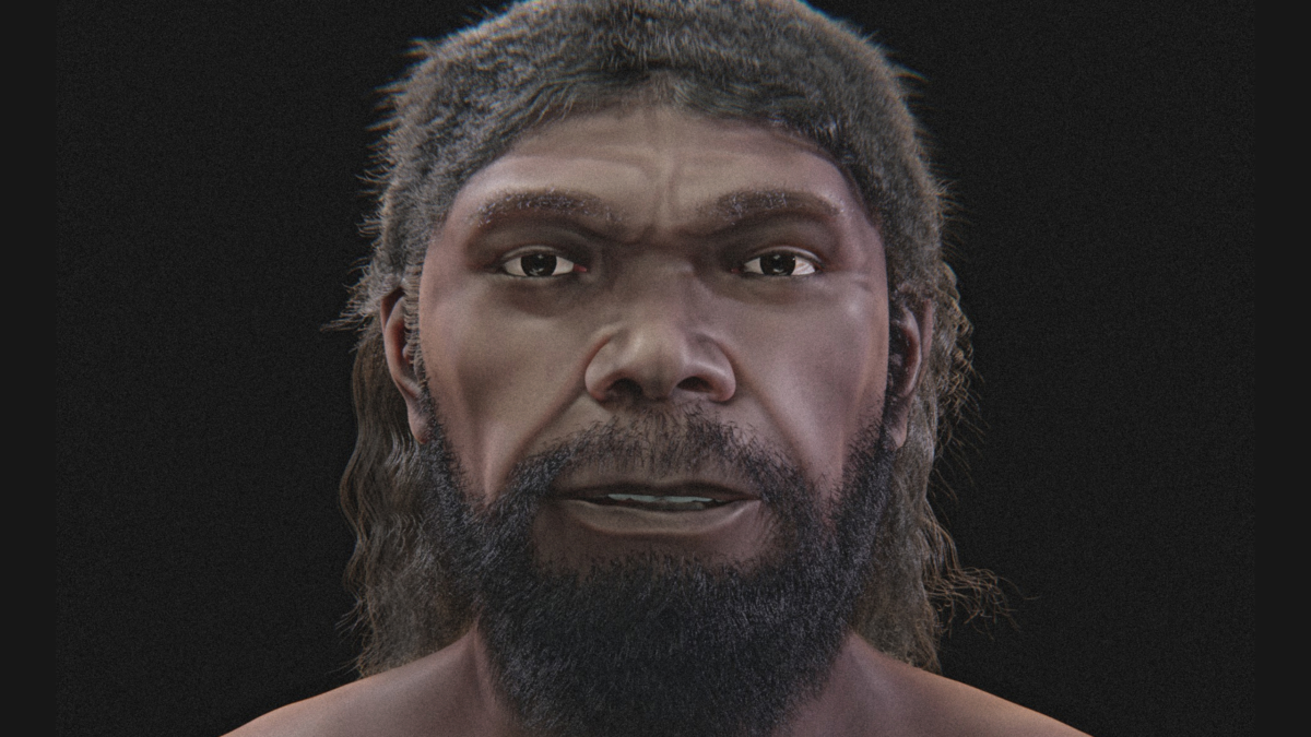
Scientists have used human fossils from Morocco to create a digital face from around 315,000 years ago, offering a glimpse of what some of the earliest known members of our species might have looked like.
Despite its strong features and projecting jaw, the face would not seem entirely out of place today.
“As for the individual from Jebel Irhoud, he might go unnoticed in a crowd, but a closer look would reveal something that sets him apart from the rest – most notably his robust features and a projecting jaw,” 3D designer Cícero Moraes told ScienceAlert.
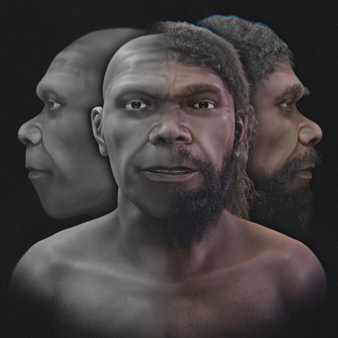 Different renderings of a face modeled from the approximately 315,000-year-old Jebel Irhoud fossils. Hair and skin pigmentation involve artistic interpretation. (Cícero Moraes)
A Skull That Rewrote Human History
The story behind that face began in the early 1960s, when a worker extracting minerals at Jebel Irhoud discovered a skull. Scientists initially thought it belonged to an African Neanderthal who lived roughly 40,000 years ago.
Later excavations uncovered more human remains and stone tools. As researchers examined the finds, their understanding of both the people and their age changed.
In 2017, two studies identified the remains as early members of our own species, Homo sapiens. Dating work placed the fossil-bearing layer at approximately 315,000 years old, with an uncertainty of about 34,000 years.
The discoveries pushed back the known history of our species by around 100,000 years, making the Jebel Irhoud remains the oldest known Homo sapiens fossils found to date. The same fossils now form the basis of the facial approximation.
Building a Face From Fragments
There was no complete skull from a single person to work with. Scientists at the Max Planck Institute for Evolutionary Anthropology had combined different fossil specimens into a more complete digital skull.
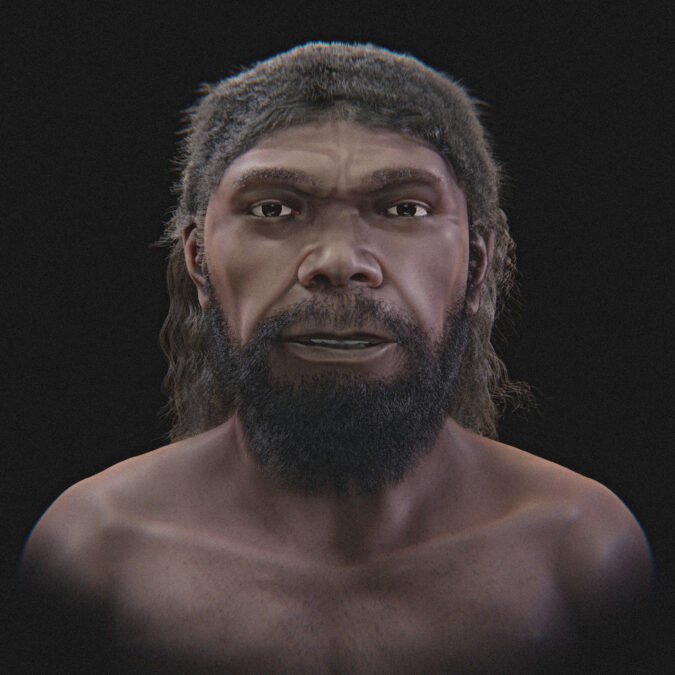 An artistic rendering of the Jebel Irhoud facial approximation. Skin pigmentation, hair, and facial hair cannot be determined directly from the fossils. (Cícero Moraes)
Most of its braincase and upper face came from the original skull, known as Irhoud 1. Missing parts, including the lower jaw and dental material, came from other remains found in the same archaeological layer. The resulting face therefore cannot be treated as an exact portrait of one individual.
Moraes considered the fit between those components convincing.
“The mandible and teeth, which belong to another individual, fit remarkably well,” Moraes said. “Therefore, even if the original mandible were found, it is very likely that the skull would not differ from its current configuration.”
His team started with images and video of the composite skull released alongside the 2017 research. They extracted video frames and used overlapping views to calculate a three-dimensional skull model, checking its scale against published measurements.
Estimating the Soft Tissue
They then needed to estimate the face covering those bones.
For this, they used a CT scan of an anonymous modern adult, which showed the skull and surrounding tissues. On the computer, they reshaped the donor’s skull to match the ancient model. The facial tissues changed with it, providing a starting point for the appearance.
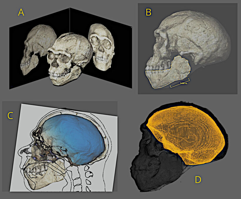 Images showing how the team reconstructed the skull digitally and modeled the space that once held the brain. Cícero Moraes; skull imagery: (Philipp Gunz, MPI EVA Leipzig / CC BY-SA 2.0)
Measurements of tissue thickness in living people helped refine the result, using the free, open-source software Blender and its OrtogOnBlender tools. This isn’t a one-off technique invented for this skull — the same anatomical deformation method has been developed and tested since at least 2013.
The bones still do most of the work. No skin or muscle survives from these ancient people to check the final face against, and the soft-tissue thickness data have to come from modern humans — a necessary stand-in, not a claim that ancient and modern anatomy are the same.
The researchers also checked the skull model’s internal dimensions. Its cranial cavity measured 1,364 milliliters, close to an earlier estimate of 1,375 milliliters calculated from CT data of the fossil. That agreement supports the skull model’s proportions, although it cannot establish how accurately the face has been reproduced.
Artistic Choices and Scientific Limits
The finished images come in two forms: grayscale renderings without hair, and a colored version with skin pigmentation, hair, and facial hair.
“Regarding subjectivity, it is fair to say that the colorized image, complete with facial hair, is the most artistic version and contains fewer scientifically robust features,” Moraes explained.
Those details cannot be read directly from the fossils. The grayscale images leave them out so viewers can examine the estimated facial shape.
Earlier analyses of the fossils identified a modern-looking face alongside an archaic-looking braincase, suggesting that the features of present-day humans developed at different times. For Moraes, one of the most anticipated moments in the entire process comes before the finishing touches, when “the basic features first emerge on the computer screen.”
The research has been published in Heritage.